|
Leg bone fracture detection using x-ray images
Johnperera [3 posts] |
16 year
|
am doing a project on leg bone (Tibia) fracture detection using image processing. I am using digital x-Ray bone fracture images to deal with this project. I was not able to find a proper method to identify bone fractures on above mentioned bone due to the high noise and variations of the fracture types.
and the color of the bone area is not identical each and every image. so its really hard to find a global threshold value for all images. So plzzz suggest me a proper segmenting and fracture detection method to move on this project....
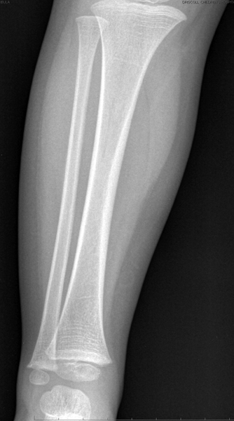
Does anyone have any idea how to handle this project using x-ray bone fracture images??? i have attached an image of that bone.
Thankx..
|
|
 |
Richard Harris from United Kingdom [6 posts] |
16 year
|
Hi,
I dont see any attached image... let me know.
|
|
|
|
Johnperera [3 posts] |
16 year
|
i hv uploaded, let me do it again. hope this works. image size sld be less than 300k to uplod
 
 brk1.zip brk1.zip
|
|
|
|
Johnperera [3 posts] |
16 year
|
its really difficult to extract features to segment the bone. Plzzzzz hlp me to figure out this.
is it possible to detect fracture area without segmenting the bone from x-ray???
|
|
|
|
Richard Harris from United Kingdom [6 posts] |
16 year
|
Hi,
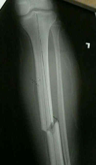
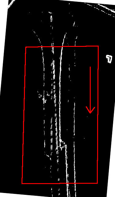
I assume the first pic is a none fracture and second is the fracture, ok the way I would approach is to apply an edge detect and then apply thresholding, this give you the below image, but I would first re-orientate the image so the leg bone faces in one direct (up - down/ left - right) then you can simple scan along the image and look for deviations from a straight path, as you can see the part with the break will be the only part within the center box that is not more or less and straight line.
BTW your image is very poor quality.
This image has prewitt applied and then thresholding... but simply have all input images orientated so the bone is at a straight line and will be easy to implement a function to scan along and look for deviations.
Let me know if have any more questions.

|
|
|
|
Johnperera [3 posts] |
16 year
|
Hi,
Plzz download brk1.zip file. I have some doubts to clarify on this sir.
1.) Do i need a global thresholding for all the images?
2.) To check deviations from a straight path, i should read the boarder of the bone pixels right??
3.) according to brk1.jpg which i have uploaded here, there is no deviation form the straight line. But there is a gap between the crack and the bone. So is that method capable for cater this fracture identification?
The thresholding values are depend on the images. so could you please let me know how to handle this problem. Also as i feel the deviation of straight line method works on some images. So could you plzz let me know how to check the fracture existence on this case :)
thankx for helping me..
brk1.zip
|
|
|
|
Richard Harris from United Kingdom [6 posts] |
16 year
|
Hi,
ok I dont know what language etc your using but something simple should be possible for this need,
you could take the image, apply prewitt, then threshold (using a value calculated by some algorithm or a fix value depending on the consistency of your images) and then line thinning (like Zhan Suen) this will give nice single pixel lines. Now assuming we orientated all our images so they are in one direction of bone, then we can scan the image by finding a bone pixel and then following the path, now if the path stops we simply scan forward so many pixels and if we happen to a pixel that is bone we know there is a break in the line (thus our fracture). For the other case where a break is a deviation that is the bone is still touching but is miss aligned then we simple check for a deviation in the line, that is it has pixels to the right or left.... and is not straight.
You happen to have IRC or something and we can chat...
The thesholding depends on what your doing, are all your images digitally acquired, or are the scanned for instance, I would guess that if you don't have consistency then some calculated threshold would be required.
|
|
|
|
johnperera [1 posts] |
16 year
|
Hii,
i'm using skype or gmail, if you prefer that application let me know..:)
Then i can send you the tested images..:)
|
|
|
|
Richard Harris from United Kingdom [6 posts] |
16 year
|
Hi, I have all skype, gmail, msn, irc, etc etc, just dont want to give my id out over on a forum....
Send your skype or gmail to this address (is secure no one can access it and stops me getting loads of spam). M8R-q6db8i@mailinator.com
|
|
|
|
Johnperera [3 posts] |
16 year
|
|
|
 |
GBB from United States [3 posts] |
15 year
|
|
This is an interesting problem. Are you doing it as an undergraduate Biomed Engineer? It would help if I understood what your real goals are.
|
|
|
|
Johnperera87 from Sri Lanka [6 posts] |
15 year
|
I'm an Undergraduate student in software engineering. For my Final yr project I'm doing a medical image processing project (Scope is to detect leg bone fractures).
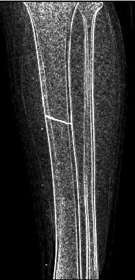
As i think I'm done with bone pre-processing part. What I'm Currently trying is, to identify the bone fracture area. I have Uploaded a processed bone image to give better idea on this. If you have any idea to detect bone fracture area and different fracture types( I'm trying to detect at least 3 types), keep replying on this thread.
Regards.

|
|
|
|
GBB from United States [3 posts] |
15 year
|
Thanks for the information. As you may know it takes physician at least about 7 years of post graduate study and thousands of examples to learn to read x rays. So given that simple fact we know that you are not going to be develop a general solution to the problem. Your problem space seems well defined (the set of three images).
I believe you asked about this problem on this message board as well. http://www.edaboard.com/thread185464.html. Both Mr. Harris and Jack have put you onto the correct path. Are you having difficulty with the conceptional aspect of the solution or it's implementation?
You have the answer to your problem. Just work with it and you will be able to show "your" solution.
The skills you gain working the problem are far more valuable than the answer to the question. That is why you where given the problem.
I for one would be very interested in hearing about your solution and lessons learned in this project.
Thanks.
GBB
|
|
|
|
Johnperera87 from Sri Lanka [6 posts] |
15 year
|
of course, Mr. Harris and Jack gave me their valuable ideas on this topic. The problem is the different type of fracture images. So one method is not capable for detecting another fracture type. I'm looking for some global method to cater above mentioned 3 fracture type images.
Mr. Harris still guiding me on this. But reducing image noise is the most difficult part. therefore I'm looking for special noise reduction method rather than well known filters.
Also Fracture pixels and noise pixels colors are almost same. That is the most difficult part in this. If there is a proper method to detect the boundary of the bone and the fracture line, it will be little bit easy for me to move on that.
Regards
|
|
|
|
GBB from United States [3 posts] |
15 year
|
Ok. in a real xray there is no noise. There is information generated by the target (the body part).There are artifacts( diffraction from some element in the target). This is important, when we read an x ray each pixel means something, two white pixels in the wrong place could be cancer. Hazy white patches in the muscle could be old blood. So if you have a nosy image give it back because it is an unacceptable x ray. Your images are good.
When humans read an x ray we look to see if something does not look normal. We compare a 2 dimensional image against a 3 dimensional model in our minds. You can't do that in this solution. That tells us that we will not be able to diagnose fractures because you do not have enough information. So what can you do?
You can identify sections of the image that are possible fractures. You will get some false positives especially near joints,but it will be a useful solution.
Your images are primarily linear with most of the vectors headed in one direction. Vectors that head about 90 to 45 degrees away from the main vectors are suspicious.
So you pre-process the images to get some good binary edges. If each pixel is important why not look at each pixel in the target area and see what its neighbors look like? Unexpected discontinuity is a flag.
I do not know your name nor do I understand how you are approaching this problem. It may help the community and you if you took some time to explain in detail your conceptional approach to this problem.
|
|
|
|
Johnperera87 from Sri Lanka [6 posts] |
15 year
|
Hii all,
I have applied sobel horizontal edge detection for the images using some free software. It was able to detect horizontal line (hairlines) on the bone. Then i was trying implementing that algorithm using C#. but the it gives so many line than the that software did. So i tried to remove that noise pixels using filter. But it destroyed the image (all the horizontal lines).
Any idea on this? Are there any problem with implementing that algo in C#?? Any C# code for this??
regards.,
|
|
|
|
[29 posts] |
15 year
|
What "free software" did you use for edge detection?
Do you have the raw data file of the image?
Scanning pixels on a 'translated' x-ray picture will induce pixel anomalies; contrast, color, resolution, size, etc. Plus the original picture is VERY large in size due to the required resolution for interpretation. Let me know if you have the original or where I can download a similar image to interrogate your needs.
|
|
|
|
Johnperera87 from Sri Lanka [6 posts] |
15 year
|
http://www.roborealm.com/uploads/21392_1.JPG.
I wanna detect horizontal lines from this image. IPLab and GIMP the software which i have mentioned as free softwares. If you can provide some C# code it will be a great help for me. Because my code detects noise pixels as well.
Regards
|
|
|
|
Elisabeth Sneha George from India [0 posts] |
15 year
|
Sir
I am doing my project on automated detection and classification of bone fracture.I need some support and help.My project submission date is on next month 14th.But it is not ready.Plz send some suggestions and codes for my project
Regards
Elisabeth Sneha George
|
|
|
|
tasneem ghnaimat from Jordan [4 posts] |
15 year
|
Hi,
Please can any one help me in my automated bone fracture detection project:
1- Preprocessing of x-ray image, and whether I should convert into binary?
2- please I cannot Explain my issue via Internet, can we communicate??!!
Any Advice,,,
Tasneem Ghnaimat
|
|
|
|
Anonymous |
15 year
|
Tasneem,
You should convert it into binary if you need to perform blob analysis i.e. analysis that would include statistics about the shape, area, etc of the object. This is normally the case in most projects that require stats but not always.
If you are using RR in your project and cannot post images or explain in detail what you need you are welcome to use our contact form to send us more specific information. Note, however, we do not respond to requests such as "I need source code for X".
Please also have example images of what you are trying to process ready to send too.
STeven.
|
|
|
|
johnperera87 from Sri Lanka [1 posts] |
15 year
|
Hi,
Can you explain me which part of the bone that u a going to consider ??... Because, one method cannot be applied for each and every bone. You have to scope down your project... And explain more then I can help u on this for some extent.
Small advice:- before starting coding, u better define your scope and make sure that your have good image collections.
Regards,
Johnperera87
|
|
|
|
tasneem ghnaimat from Jordan [4 posts] |
15 year
|
Hi,
Ok. I will work on long bones, and specifically (leg or hand bone), but this will be determined according to the availability of data
Please I need help from you, if u r willing to help m, please tell me to ask u some questions for clarification purpoes,
Tasneem
|
|
|
|
Johnperera87 from Sri Lanka [6 posts] |
15 year
|
Hi,
Ya sure, Feel free to ask any question...... You better collect good dataset, then you will be able to identify the patterns of the fractures... after that move on thinking suitable methods for this.
Regards
Aruna
|
|
|
|
tasneem ghnaimat from Jordan [4 posts] |
15 year
|
Ok.
I read about Statistical models (Active Appearance model and Active shape model), will they help me in bone detection, or it is better to use image processing techniques, like sobel filter, gaussian,etc.
Can u provide me with an email that I can contact u on it? I have lot of questions,,, Please
|
|
|
|
Johnperera87 from Sri Lanka [6 posts] |
15 year
|
Hi,
It depends....But image processing techniques will make your job easy. Such as edge sharpening, contrast / brightness, Edge Detection, alot more... here is my email : johnperera87@gmail.com
|
|